The diagnosis your doctor may be missing
Picture this: you have been dealing with a persistent sinus infection for months. You have tried multiple
courses of antibiotics, nasal sprays and antihistamines. Your ENT specialist confirms that your maxillary
sinus is inflamed, but cannot pinpoint a clear cause. The headaches, facial pressure, and congestion keep
coming back. What nobody has asked you yet is: how are your upper back teeth?
This scenario plays out more often than most people realize — in Costa Rica and globally. Odontogenic
maxillary sinusitis, also known as Maxillary Sinusitis of Endodontic Origin (MSEO), is one of the most
underdiagnosed conditions in routine medical practice. The source of the problem is not in the nose or the
sinus cavity itself — it is inside an infected tooth. And the specialist who can solve it is an endodontist.
1. How common is dental-origin sinusitis?
The numbers are striking. The American Association of Endodontists (AAE) — one of the most authoritative dental specialty organizations in the world — issued an official Position Statement in 2018 formally recognizing MSEO as a distinct clinical entity requiring specialized management. According to that document and the supporting body of research, more than 40% of all maxillary sinusitis cases have an odontogenic origin, meaning a tooth is responsible.
That figure becomes even more telling when a specific presentation is considered: unilateral sinusitis — inflammation affecting only one sinus. In those cases, the probability of a dental cause rises above 70%. Despite this, a systematic review of over 85 clinical guidelines for sinusitis management found that only three recommended a dental evaluation, and none suggested referral to an endodontist.
“Studies indicate that more than 40% of maxillary sinusitis cases are odontogenic in origin, increasing to over 70% when maxillary sinus infections are unilateral.” — American Association of Endodontists (AAE), Position Statement, 2018
In Costa Rica, where many patients defer dental treatment for extended periods, the number of untreated tooth infections that progress to sinus involvement is considerable. At DensCare in Escazu, Dr. Tiapa regularly receives patients who have undergone months of medical treatment for sinusitis without resolution — and whose CBCT imaging reveals the real culprit: a necrotic upper molar.
2. The anatomy behind the problem: how a tooth infects the sinus
Understanding the connection between tooth infections and sinus disease requires a brief anatomy lesson. The maxillary sinus is an air-filled cavity within the upper jaw, and its floor sits in very close proximity to — and sometimes in direct contact with — the roots of the upper molars and premolars. In many patients, the roots of the upper first molar literally reach into or through the sinus floor.
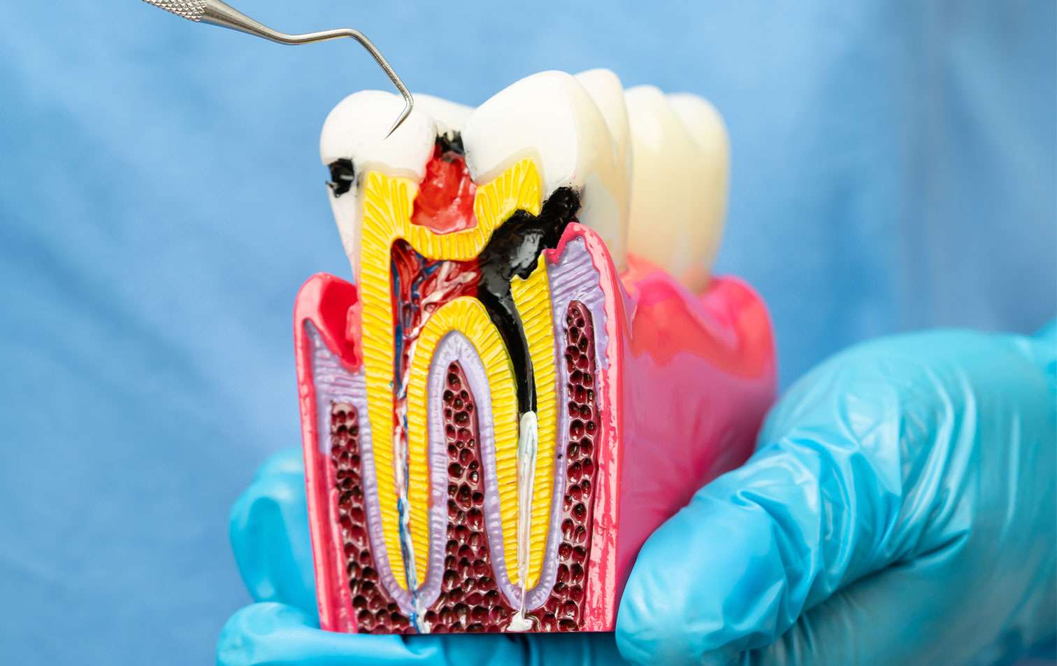
When those roots develop a dental infection — through deep decay, dental trauma or a failing previous treatment — the tooth’s pulp dies and becomes a reservoir of anaerobic bacteria. Over time, this infection destroys the surrounding bone, perforates the sinus floor, and seeds the sinus cavity with oral pathogens. The result is a chronic inflammatory response: odontogenic maxillary sinusitis.
Unlike viral or allergic sinusitis, the odontogenic form does not respond to antibiotics or nasal treatments because the infectious source — the diseased tooth — remains active. The sinus can be surgically cleaned, but if the dental infection is not addressed, the sinusitis will inevitably return.

Dr. Tiapa’s case — DensCare, Costa Rica | CBCT coronal view. BEFORE (left): Perforation of the maxillary sinus floor by an
apical lesion with fluid-filled cavity. AFTER (right): 6-month follow-up. Maxillary sinus fully clear of inflammatory changes
following root canal treatment.
3. The right diagnosis: cbct imaging and the microscope
Accurate diagnosis of tooth-related sinus disease demands advanced imaging technology. Standard periapical X-rays, while useful, frequently underestimate the actual extent of periapical lesions and may not show sinus involvement at all. The current gold standard is cone beam computed tomography (CBCT), which provides a three-dimensional view of the relationship between periapical pathology, surrounding bone, and the maxillary sinus.
At DensCare, Dr. Tiapa performs detailed CBCT analysis to identify: periapical lesions in roots adjacent to the sinus, integrity of the sinus floor, degree of mucosal thickening, and extent of the infection. This three dimensional assessment is not possible with conventional dental X-rays — and it is precisely why so many of these cases go undetected in general medical practice.
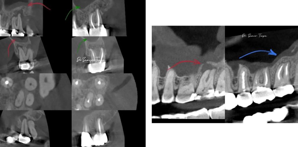
Dr. Tiapa’s cases — DensCare, Costa Rica. Left: Multi-view CBCT series. Red arrows: active periapical lesion before
treatment. Green arrows: complete resolution post-treatment. Right: Pre/post comparison — periapical bone loss fully resolved following root canal therapy.
4. The right treatment: why you need an endodontist, not an ent
This is where a critical and costly mistake is often made: treating the sinus without treating the tooth.
Research shows that functional endoscopic sinus surgery (FESS) performed without addressing the dental source carries a failure rate exceeding 40%. Patients experience temporary relief, but sinusitis returns because the original infectious source — the diseased tooth — is still present.
An endodontist is the dental specialist specifically trained to diagnose and eliminate pulpal and periapical infections.
Through root canal treatment, the necrotic, infected tissue inside the tooth is removed, the root canal system is thoroughly disinfected, and the tooth is sealed to prevent recontamination. By eliminating the infectious source, the patient’s immune system can then resolve the
sinus inflammation naturally — without surgery, without ongoing antibiotics.
In cases where the infection has produced a large cyst or granuloma that does not fully heal with conventional root canal therapy, an endodontist can perform apicoectomy surgery — a microsurgical procedure that directly removes the apical lesion with minimal disruption to surrounding bone and tissue.
Performed under an operating microscope, this procedure offers predictable, long-term results even in complex anatomical situations.
ENT specialists and primary care physicians are essential for managing the airways — but if your maxillary sinusitis is not responding to conventional treatment, the cause may be dental. In that case, the right specialist is an endodontist.
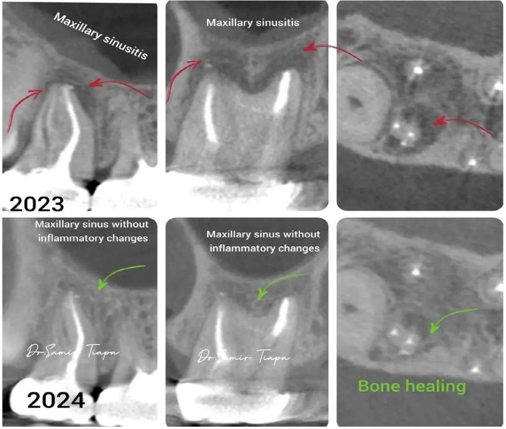
Dr. Tiapa’s case — DensCare, Costa Rica. 2023 vs 2024. Top row (red): active maxillary sinusitis with sinus opacification and periapical lesion. Bottom row (green): maxillary sinus without inflammatory changes and complete bone healing following root canal treatment.
5. Denscare: microscope endodontics in escazu, Costa Rica
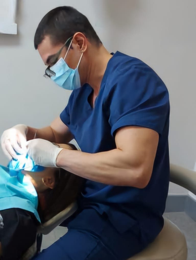
Dr. Samir Tiapa is an endodontist with over 15 years of clinical experience and a member of the Academia Costarricense de Endodoncia (Costa Rican Academy of Endodontics). At DensCare,
located at Torre Medica 2, 2nd Floor, Office 203 within the Hospital CIMA Escazu campus, he offers the highest standard of diagnosis and treatment for dental-origin sinus disease in Costa Rica — serving both
local patients and international visitors.
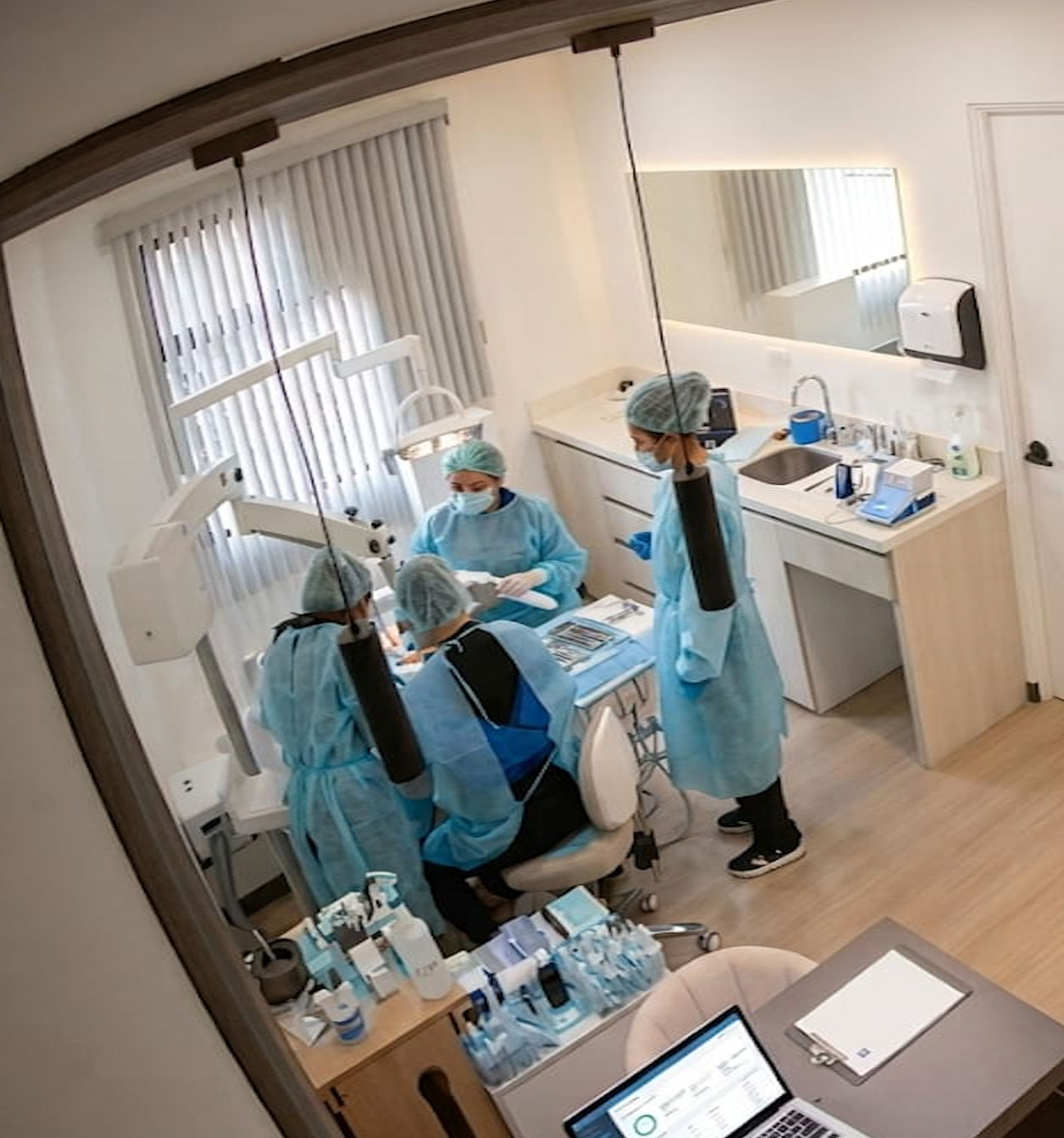
Every treatment at DensCare is performed under a dental operating microscope, providing up to 25x magnification. This allows Dr. Tiapa to identify structures invisible to the naked eye: calcified canals, root
fractures, apical isthmuses, and direct sinus communications. This level of precision translates into significantly better clinical outcomes and a higher probability of saving the natural tooth.
The diagnostic protocol at DensCare includes full CBCT analysis, pulp vitality testing, comprehensive medical history review, and interdisciplinary coordination with the patient’s physician when needed. Every
case is documented radiographically before and after treatment to provide objective confirmation of dental infection resolution and maxillary sinus recovery.
Conclusion: not all sinusitis is what it seems
Odontogenic maxillary sinusitis is a real, common and — critically — solvable condition. If you or a family
member has recurrent unilateral sinusitis, persistent facial pressure or cheek pain, or nasal discharge that
does not respond to medical treatment, we strongly recommend seeking a specialist endodontic evaluation. A CBCT scan can reveal in minutes what years of medical treatment may have failed to uncover.
Sinusitis that won’t go away? See an endodontist. DensCare — Dr. Samir Tiapa | Torre Medica 2, Office 203, Hospital CIMA Escazu, Costa Rica Tel: +506 2208-8203 | Emergency: +506 7140-0509