Why the microscope changes everything in endodontics
Imagine being asked to repair a precision watch using only your bare hands — no magnifying glass, no direct lighting, working through an opening the size of a pinhead. That was endodontics for decades: an extraordinarily delicate procedure performed, for the most part, in the dark.
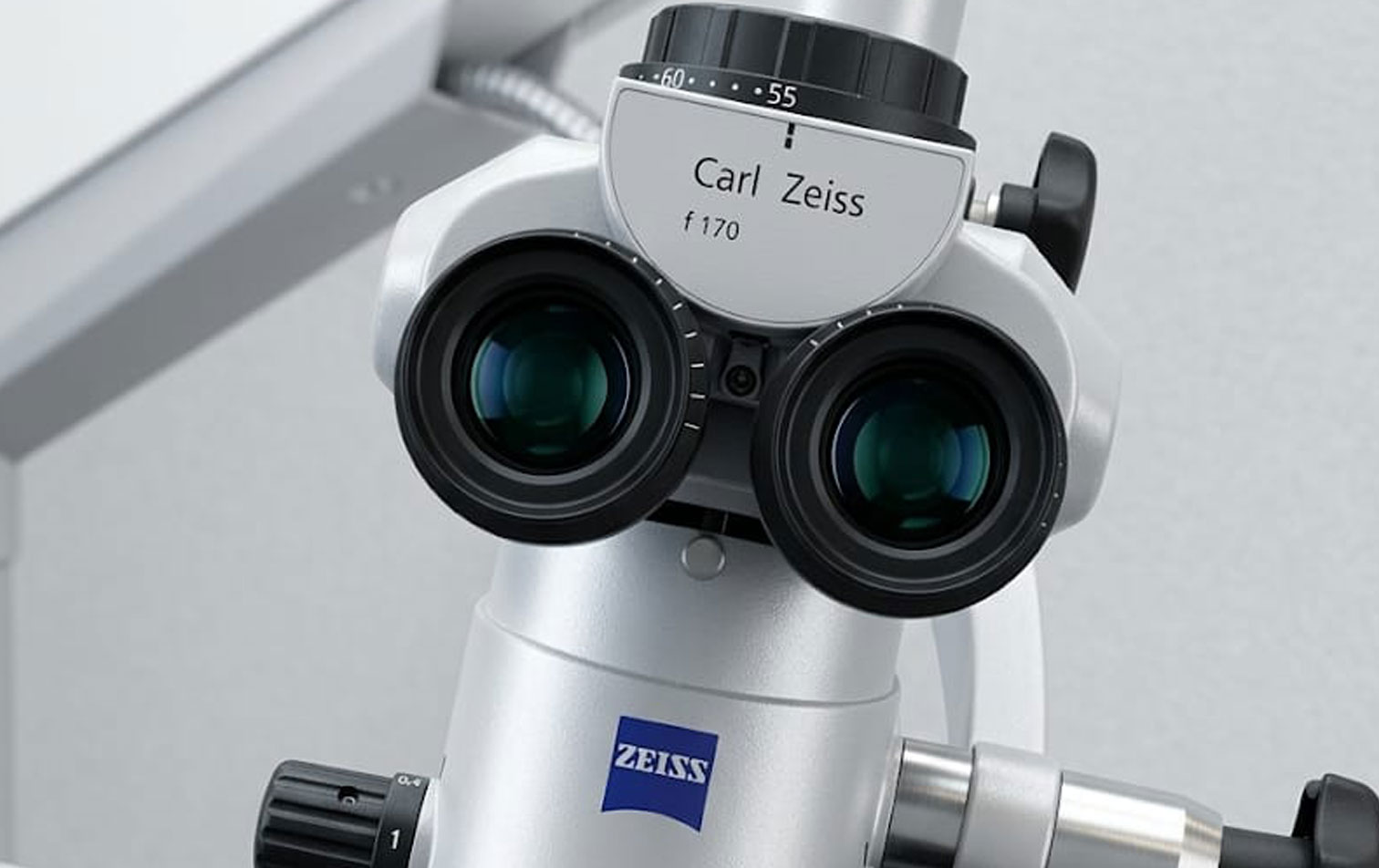
That changed. And the instrument that changed it is the surgical operating microscope.
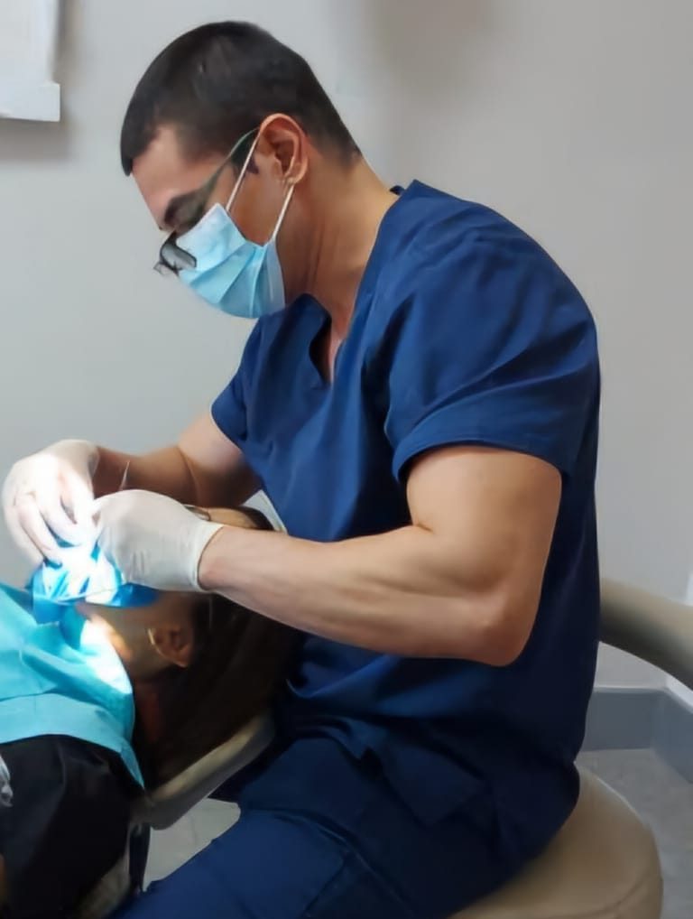
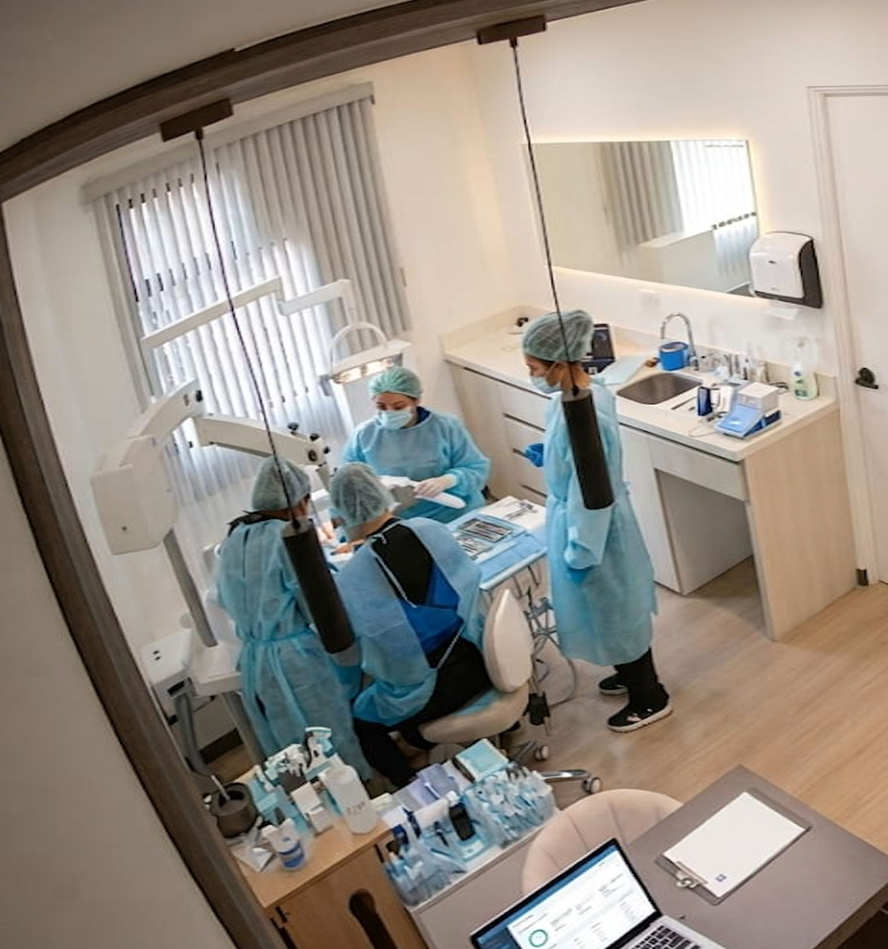
At DensCare, 100% of our treatments are performed under the microscope. This is not an aesthetic detail or a technological luxury: it is the difference between guessing and seeing. Between intuition and confirmation. Between a treatment that will probably work and one we know has been done right.
In this article, we explain why the microscope has become the gold standard in specialized endodontics — and what that means for you as a patient.
The problem with working without magnification
The root canal — the internal space of the tooth where the dental pulp lives — is a microscopic
structure. In posterior teeth, it can measure between 0.2 and 0.5 millimeters in diameter. For
reference: a human hair is approximately 0.07 millimeters wide. We are talking about spaces
that are barely visible to the naked eye.
Inside those microscopic spaces is where everything important happens: the infection causing
pain, the bacteria compromising the bone, the additional canals that — if left untreated — may
be the reason a treatment fails years later.
Without magnification, the endodontist works with what they can perceive through touch and
experience. That has value — the best specialists develop a remarkable sensitivity — but it has
clear limits. Some things simply cannot be felt: an early vertical fracture, a calcified canal that
transmits no resistance, a bifurcation in canal anatomy that changes the entire treatment plan.
The microscope eliminates that uncertainty.
What the microscope reveals that the eye cannot
With magnifications ranging from 4x to 25x, and high-intensity coaxial illumination directed straight into the operative field, the surgical operating microscope reveals anatomy that would otherwise remain hidden.
Additional canals
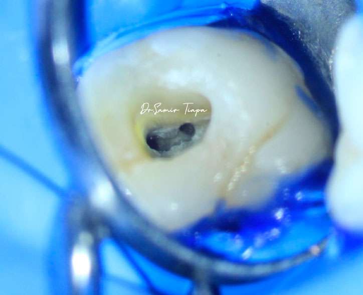
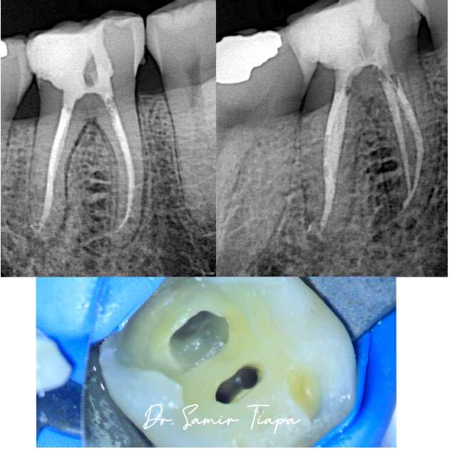
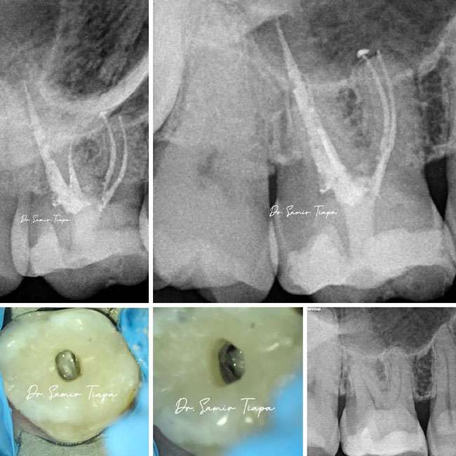
One of the most frequent findings under the microscope is the presence of canals that do not appear on conventional X-rays. The upper molar, for example, frequently has a fourth canal — the MB2 — located in a position that can only be identified visually with adequate magnification. Studies published in leading endodontic journals indicate this canal is present in more than 70% of upper first molars, yet is detected at a much lower rate when no microscope is used. That untreated canal is one of the most common causes of endodontic failure.


Cracks and fractures
Dental fractures are deceptive: they can be invisible on X-rays and go completely unnoticed by the unaided clinical eye. Under the microscope, a fracture line that compromises the prognosis of the tooth can be identified before treatment begins — critical information that prevents investing time and money in a tooth that cannot be saved.
Calcifications
“With age, or as a response to prior trauma, canals can partially calcify. Navigating through calcified tissue requires seeing exactly where you are working. The microscope allows the specialist to distinguish calcified tissue from healthy surrounding tissue, reducing the risk of perforation and preserving dental structure.”


Separated instruments
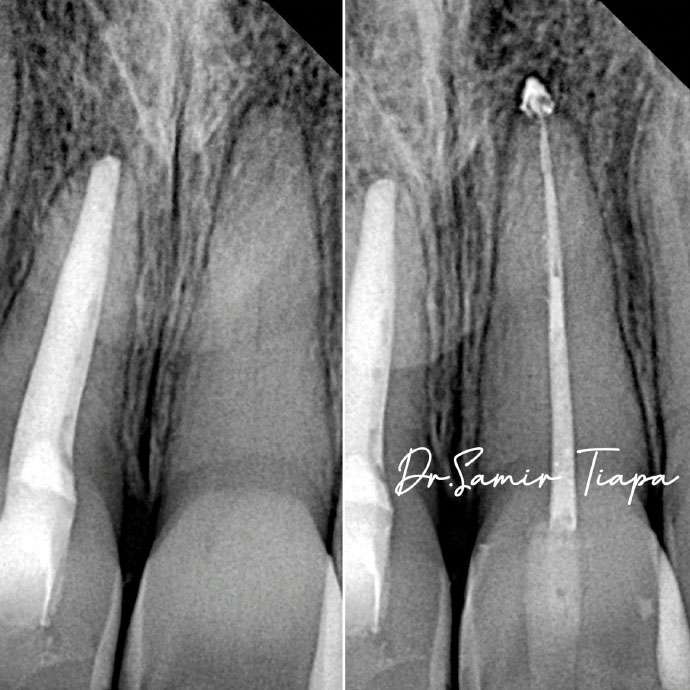
When an endodontic file breaks inside the canal — an infrequent but possible complication — managing it requires seeing exactly where it is, how it is oriented, and how much space surrounds it. Without a microscope, many of these situations end with the instrument left inside the canal. With a microscope, retrieval is possible in many cases.
Precise removal of decay
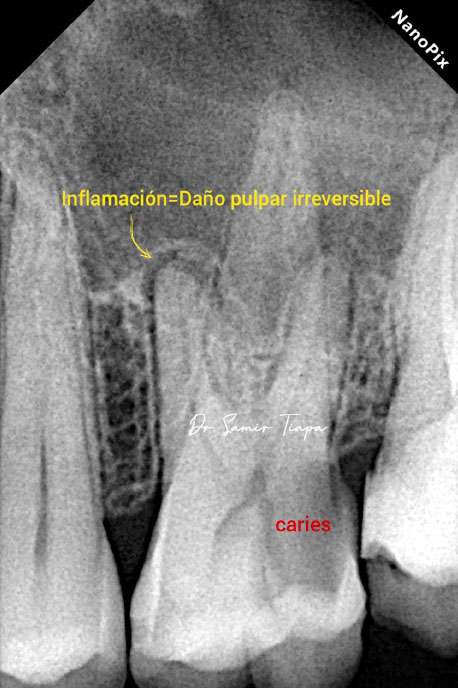
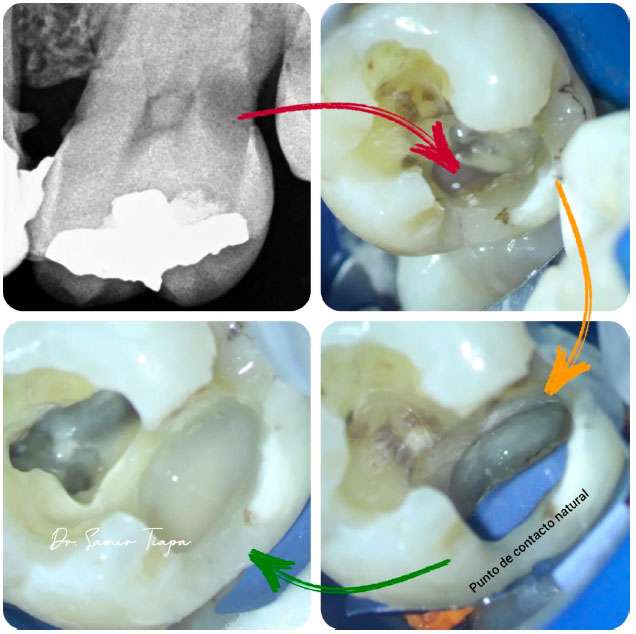
Before any endodontic treatment begins, the tooth must be free of decayed tissue. Under the microscope, this step — which might seem routine — achieves a level of precision that changes the final outcome. To the naked eye, identifying the exact boundary between infected and healthy dentin is difficult. Without magnification, the clinician tends to err in one of two directions: removing too much tissue out of caution, or leaving residual decay that compromises the seal and long-term longevity of the treatment.
Under magnification, that boundary becomes visible. Infected dentin has a different texture, color, and consistency from healthy dentin, and the microscope allows the specialist to identify that threshold with a precision that is simply not achievable any other way. In cases where decay is deep and approaches the pulp, this precision can even be the difference between needing a full root canal or achieving a successful pulp capping that preserves the tooth’s vitality.


Conservative access: less is more
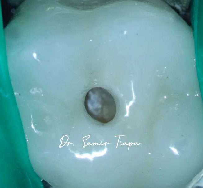
One of the most important shifts the microscope brought to modern endodontics is the philosophy of minimal access. For years, standard practice called for wide access cavities — “”opening up”” the tooth — to ensure visibility and working space. The problem is that this approach came at a cost: it weakened the coronal structure of the tooth, increased the long-term risk of fracture, and sacrificed healthy tissue that can never be recovered.
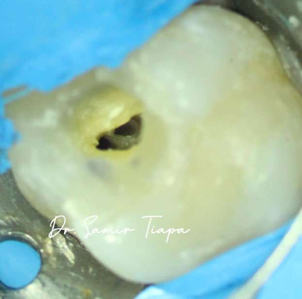
With the microscope, that logic is reversed. Magnification and direct illumination allow the specialist to work through much smaller openings, precisely because they can see what they are doing even in a confined space. The result is a tooth that retains more of its original structure: more dentin, more enamel, more mechanical strength. This approach — known as minimally invasive endodontics — is backed by evidence: teeth treated with conservative access show lower rates of vertical root fracture, one of the most serious complications and one that frequently leads to extraction.

Surgery: where the microscope becomes indispensable
If the microscope makes an important difference in conventional treatment, in periapical surgery — also called an apicoectomy — it is absolutely indispensable.
Periapical surgery involves accessing the tip of the root through the bone, removing infected tissue, and sealing the end of the root with a biocompatible material. It is a high-precision procedure in a very small operative field, typically with limited access.
Under the microscope, the surgeon can prepare the apical cavity with ultrasonic instruments measuring just millimeters, verify that the seal is complete, identify isthmuses and connections between canals that would otherwise go untreated, and visually confirm the quality of the work before closing.
Comparative studies between periapical surgery with and without a microscope are clear: success rates with the microscope are significantly higher. We are talking about moving from success rates of 40–60% in traditional surgery to over 85–90% with magnification. That translates directly into teeth saved rather than extracted.
What the microscope means for you as a patient
More conservative treatments
Seeing with precision means acting with precision. That means less tooth structure removed, smaller access openings, and greater preservation of the natural tooth.
Preservation of healthy tissue
Every millimeter of dentin preserved matters. Dentin is the tissue that gives the tooth its strength; once removed, it does not regenerate. Under the microscope, the specialist can precisely distinguish infected tissue from healthy adjacent tissue, and act only where necessary. In retreatment cases, this ability is especially valuable: removing a previous filling without sacrificing unnecessary dentin, navigating around a post or a calcification, locating the original canal without creating false paths — all of that requires seeing. And seeing well.
More accurate diagnoses
Before any treatment begins, the microscope enables an evaluation that goes beyond what an X-ray reveals. This leads to better-founded treatment plans and fewer surprises during the procedure.
Fewer retreatments
A treatment done correctly from the start — with all canals identified and treated, with a seal visually verified — is far less likely to require intervention again in the future.
Greater safety in complex cases
In cases of unusual anatomy, severe calcifications, or teeth with a complicated history, the microscope is not a luxury: it is what makes treatment possible.
Specialized endodontics: more than a title
In Costa Rica, as in many countries, any general dentist can perform a root canal. And many do it well, within the limits of straightforward cases with conventional anatomy.
But specialized endodontics exists precisely because some cases require more: more training, more experience, better tools, and the clinical judgment to recognize when a case exceeds what can be resolved without a microscope, without CBCT imaging, without endodontic ultrasound.
The endodontic specialist has dedicated additional years of training exclusively to this field. They know root anatomy in its most complex variants. They have managed complications that a general dentist will rarely encounter. And they work with tools — among them the microscope
— built for that level of demand.
At DensCare, every treatment — from a dental emergency to a complex periapical surgery — is performed under the surgical operating microscope, with rigorous aseptic protocols and the time required to do it right.
An investment in the tooth you already have
Preserving a natural tooth will always be preferable to replacing it. A dental implant is an excellent solution when there is no other option — but it does not fully replicate the function, feel, or biological integration of a natural tooth.
The microscope, in the context of specialized endodontics, is precisely that: a tool to maximize the chances of keeping what you already have.
See more. Work better. Save more teeth.
That is what the microscope does. And that is why at DensCare we cannot”
“conceive of endodontics any other way.