If this sounds familiar
You had an implant placed in a front tooth, one that shows when you smile. You invested time, money and patience. And still, the implant didn’t work.
Maybe you were told there is “no bone left,” that it can’t be tried again, or that you’ll have to settle for a removable denture. Many patients come to us exactly like this: tired, uncertain and afraid of going through it all again.
This case is for you.
The starting point
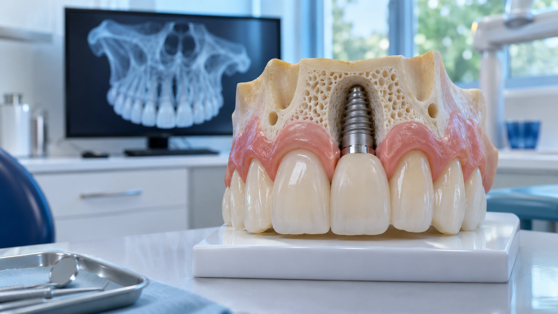
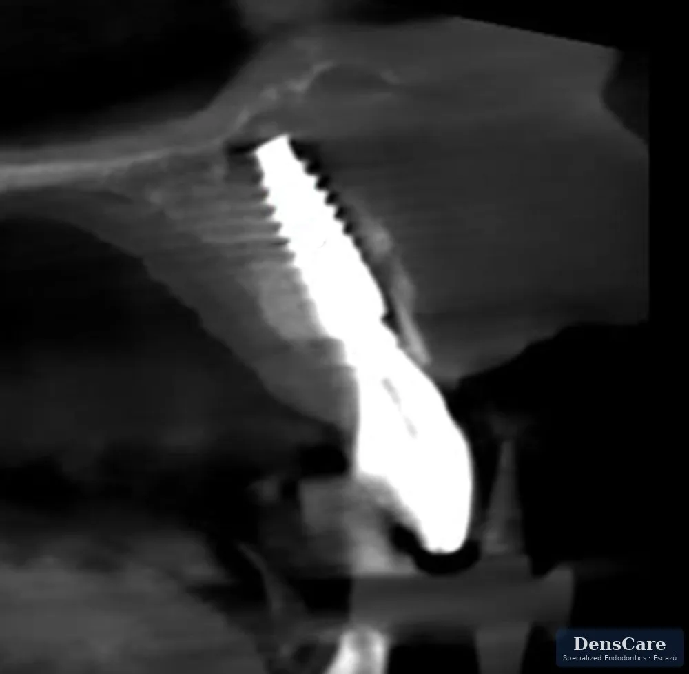
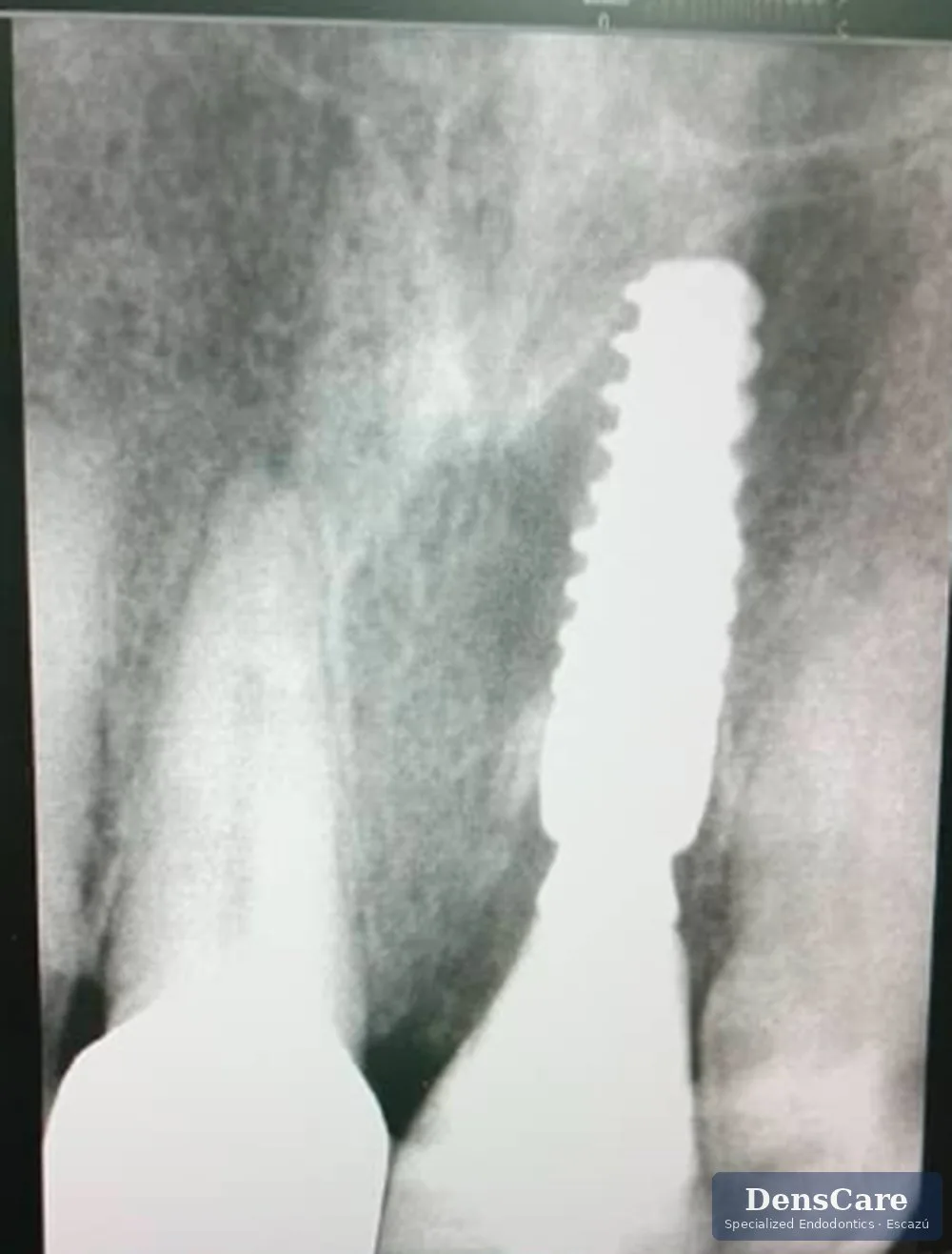
This is how the implant looked before it was removed. At first glance it seems to be in place, but an implant can look “well placed” and still not be healthy: what determines its success is the bone around it and what was left underneath.
What we found
A patient came to DensCare after his implant in the upper left central incisor failed at another facility. Before doing anything, we did the most important thing: understand why it failed.
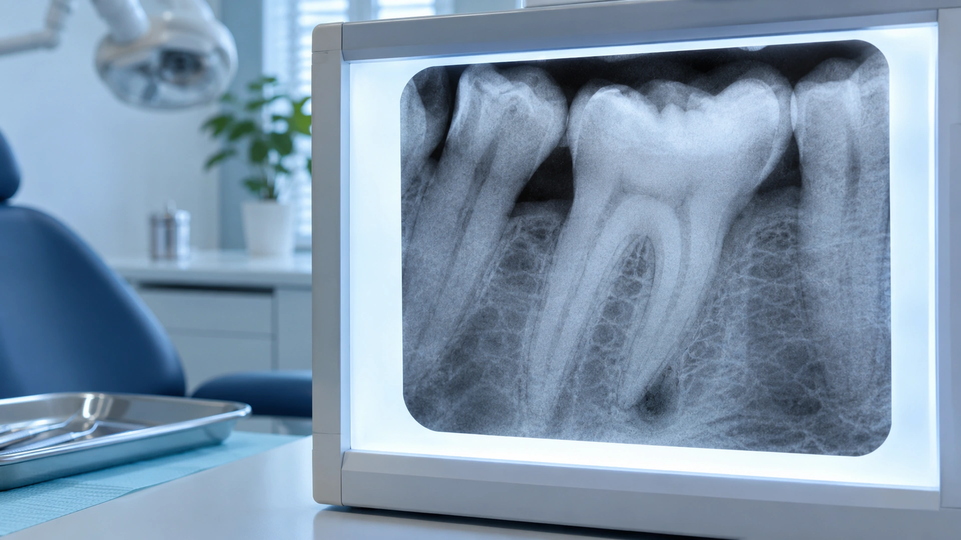
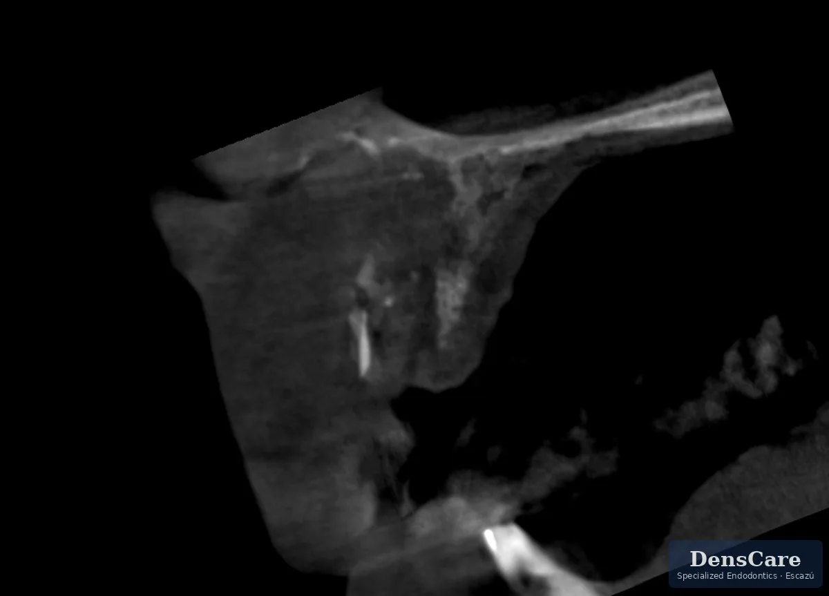
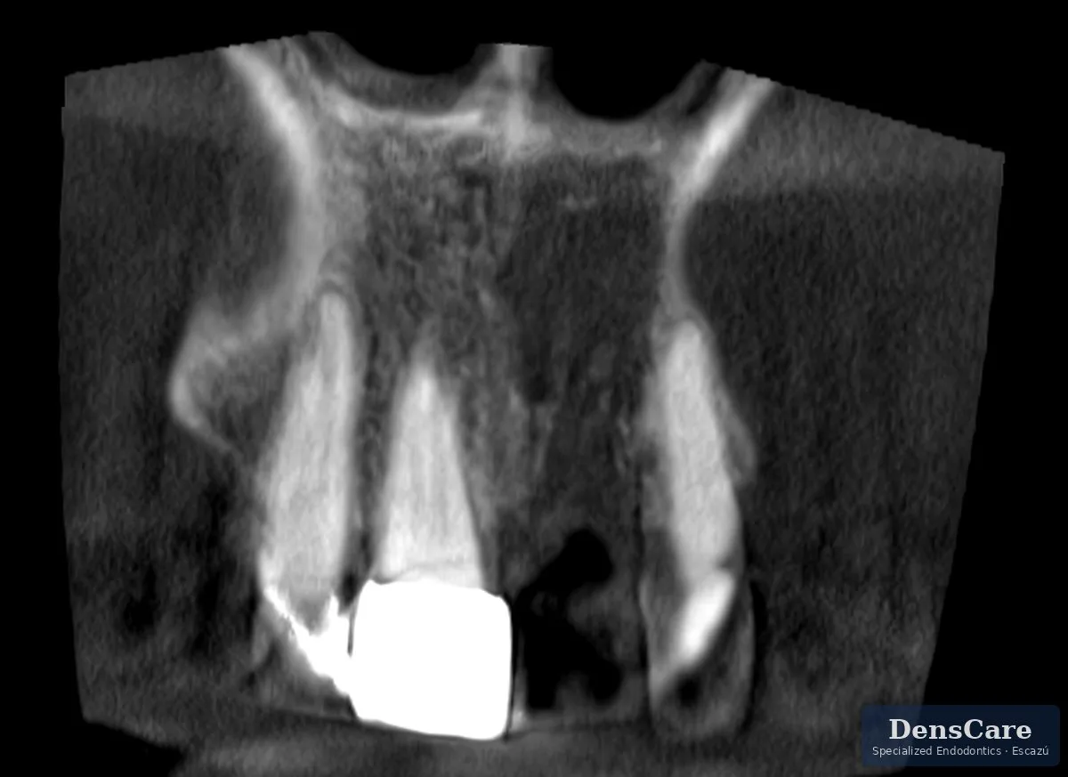
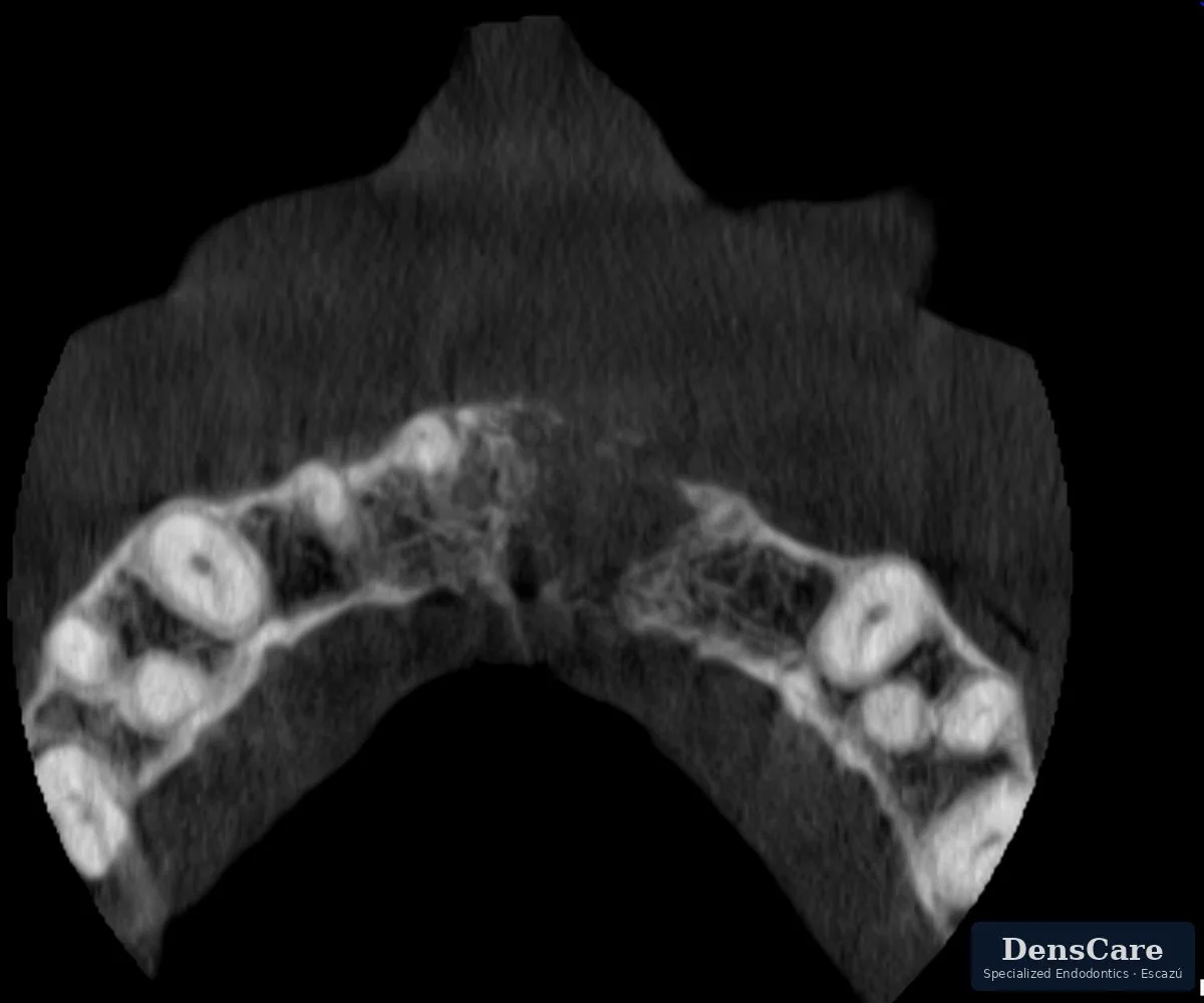
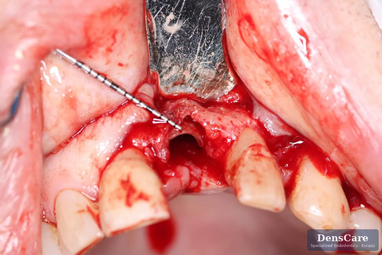
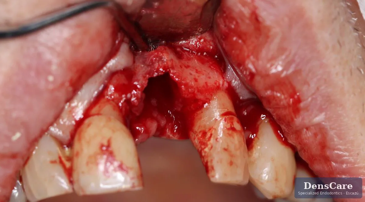
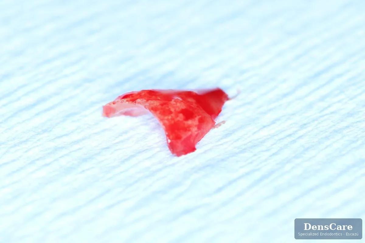
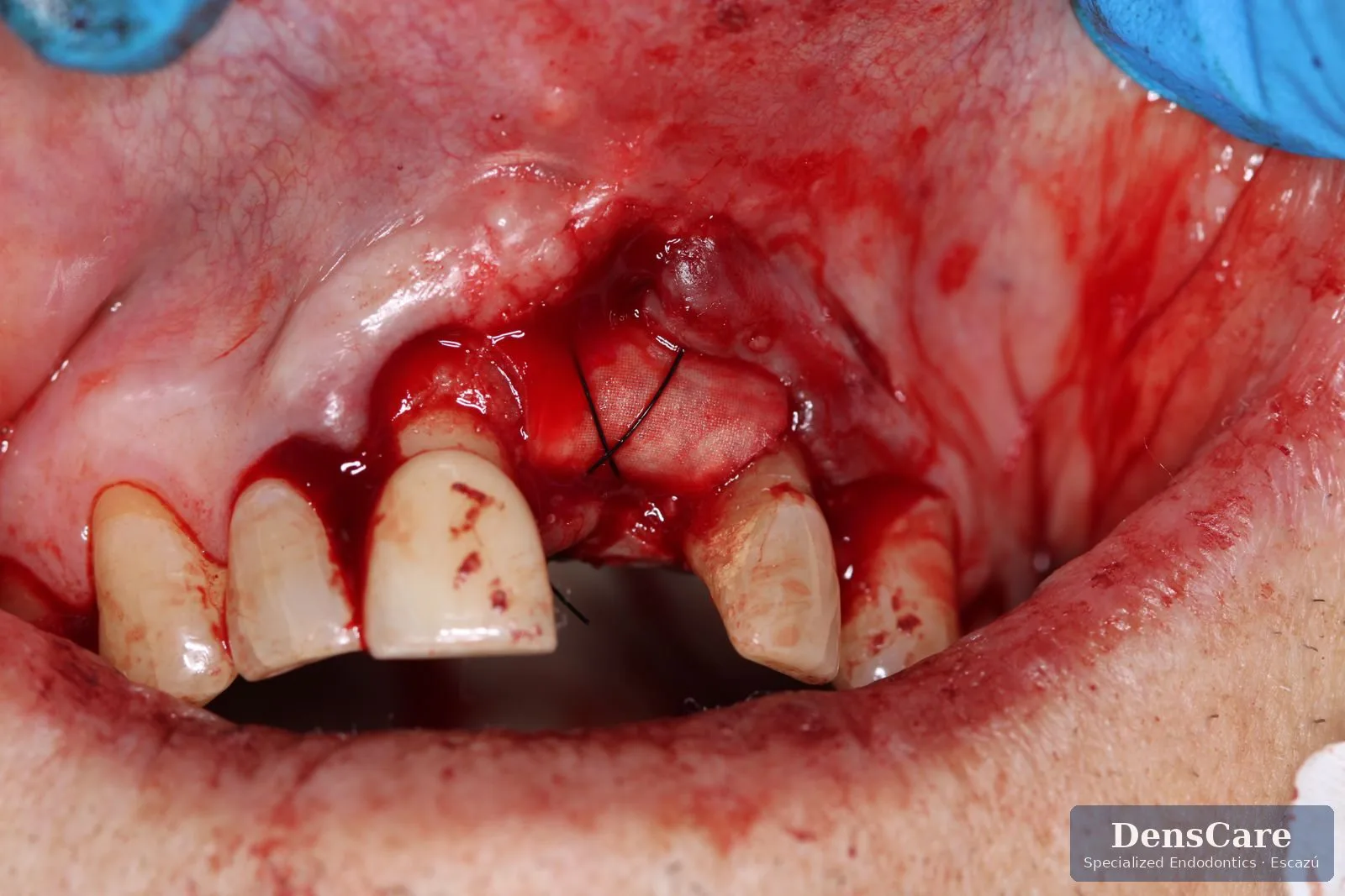
The 3D scan showed a bone defect nearly 18 mm deep. When we opened the site, the cause appeared: fractured bone fragments and root remnants never removed during the original extraction. No implant can hold on ground like that. The problem wasn’t the patient or his bone; it was what had been left inside.
What we did
- Fully removed the root remnants and damaged bone fragments: the cause of the failure.
- Cleaned the defect and filled it with a bone graft prepared with an antibiotic.
- Protected it with a collagen membrane that the body absorbs on its own.
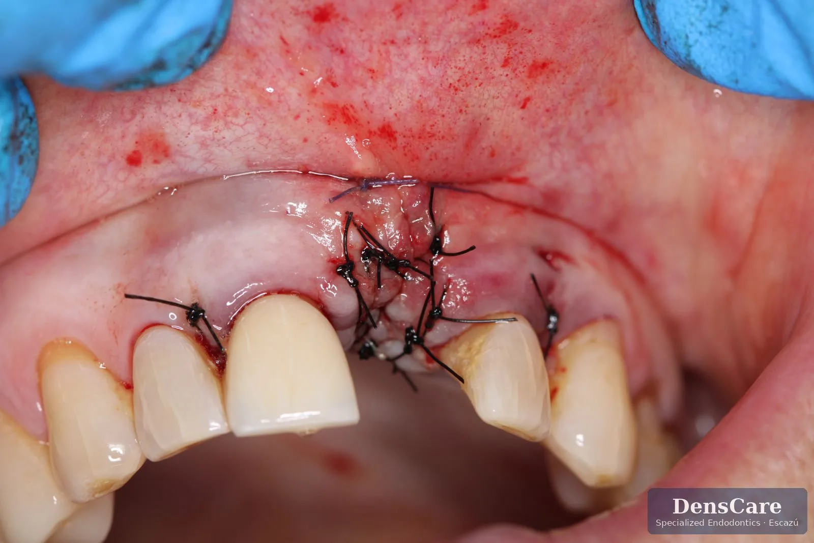
- Closed the gum with stitches so new bone can form undisturbed.
Case record
What comes next
What you should know
- A failed implant almost always has a cause. Finding it is the first step to not repeating the mistake.
- “No bone left” isn’t always final. In many cases, lost bone can be rebuilt.
- We never try again blindly. At DensCare, every case starts with a 3D scan and ends with documented follow-up.
Images published with authorization. Patient details omitted to protect privacy. Every case is unique and requires individual evaluation.